
Understanding the Role of Ovaries in Women’s Health
The ovaries are among the most important organs in the female reproductive system. These two small, almond-shaped structures are located on either side of the uterus and perform functions that directly influence fertility, menstruation, pregnancy, and hormone production. Although they are relatively small in size, their role in maintaining reproductive health is enormous. Every month, the ovaries prepare and release an egg while producing essential hormones such as estrogen and progesterone that regulate the menstrual cycle.
Many women become concerned when they receive an ultrasound report showing ovarian measurements. Seeing numbers, dimensions, and medical terminology can be confusing, especially if you are trying to conceive or experiencing irregular periods. Understanding what constitutes a normal ovary size can help reduce unnecessary anxiety and provide a clearer picture of your reproductive health.
Healthcare professionals often evaluate ovarian size as part of routine gynecological examinations, fertility assessments, and investigations for conditions such as PCOS, ovarian cysts, and hormonal disorders. However, ovary size alone does not determine fertility or overall reproductive health. It is only one part of a much larger clinical picture that includes hormone levels, follicle count, menstrual regularity, and symptoms.
What Is the Normal Ovary Size?
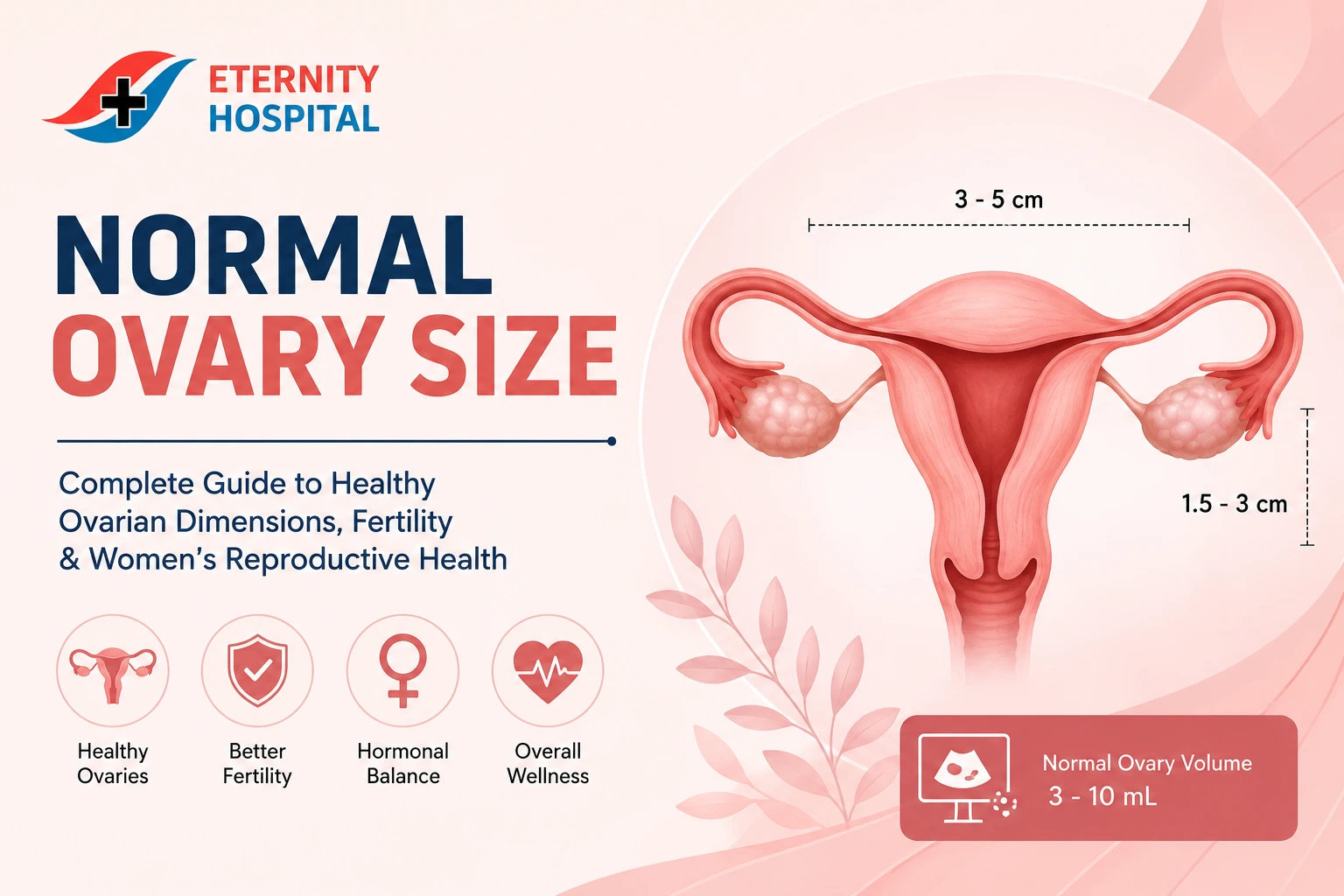
A common question women ask after an ultrasound examination is, “Are my ovaries normal?” The answer depends on age, reproductive status, and individual health factors. In general, healthy ovaries in women of reproductive age fall within a recognized range of dimensions. Current medical references suggest that the normal ovary size in adult women typically measures approximately 3 to 5 centimeters in length, 1.5 to 3 centimeters in width, and 0.6 to 1.5 centimeters in thickness.
Standard Ovary Measurements
The following table shows typical ovarian measurements:
Measurement Normal Range
Length 3 – 5 cm
Width 1.5 – 3 cm
Thickness 0.6 – 1.5 cm
Volume 3 – 10 mL
These measurements are considered average ranges and may vary from woman to woman. One ovary measuring 4.2 cm and another measuring 3.8 cm can both be completely normal. Doctors are generally more interested in ovarian structure, follicle count, and blood flow rather than focusing solely on size.
Normal Ovarian Volume
Ovarian volume is calculated using a specific formula that considers the ovary’s length, width, and thickness. Most healthy reproductive-age women have an ovarian volume between 3 and 10 mL. Volume often provides a better indication of ovarian health than a single-dimensional measurement because it reflects the ovary’s three-dimensional size.
Normal Ovary Size by Age
Ovarian size changes significantly throughout a woman’s life. Just as the body evolves from childhood to adulthood and eventually into menopause, the ovaries undergo similar transformations. Understanding these changes helps women interpret ultrasound findings more accurately.
Childhood and Puberty
Before puberty, the ovaries are relatively small because they are not actively producing mature eggs. Hormonal activity is minimal during childhood, which results in lower ovarian volume and smaller dimensions. As puberty approaches, hormonal signals from the brain stimulate ovarian development, leading to gradual growth.
During adolescence, follicles begin developing within the ovaries, and menstruation eventually starts. This stage marks the transition from childhood ovarian dimensions to adult reproductive sizes. Variations during puberty are common and usually reflect normal developmental processes rather than underlying health concerns.
Reproductive Years
The reproductive years represent the period when ovarian size is generally at its peak. Studies show that ovarian volume reaches its highest average levels during the late teenage years and early twenties. Healthy reproductive-age women typically have ovarian volumes ranging from 3 to 10 mL, with average values often around 6 to 8 mL.
During this phase, the ovaries are actively producing eggs and hormones. Monthly follicle development causes natural fluctuations in ovarian dimensions. Therefore, measurements taken at different times of the menstrual cycle may vary slightly without indicating any health problem.
Perimenopause and Menopause
As women approach menopause, ovarian activity gradually declines. The number of available eggs decreases, hormone production falls, and ovarian volume becomes smaller. Research indicates that ovarian volume significantly decreases after menopause due to reduced follicular activity.
Smaller ovaries in postmenopausal women are usually expected and do not necessarily indicate disease. In fact, shrinking ovarian size is a normal part of reproductive aging. Doctors interpret ovarian measurements differently depending on whether a woman is premenopausal or postmenopausal.
Right Ovary vs Left Ovary Size Differences
Many women worry when their ultrasound report shows one ovary larger than the other. Fortunately, a slight asymmetry is entirely normal. The human body is not perfectly symmetrical, and the ovaries are no exception.
Studies and clinical observations reveal that the right ovary is often slightly larger than the left ovary. This difference may occur because of variations in blood supply, anatomical positioning, and ovulation patterns. During a particular menstrual cycle, one ovary may contain a dominant follicle preparing to release an egg, making it temporarily larger than the opposite ovary.
A size difference of a few millimeters or even several centimeters is not automatically concerning. What matters more is whether the ovary maintains a normal appearance on imaging. Doctors assess factors such as cysts, masses, blood flow, and follicle distribution before determining if a size difference requires further investigation.
Women should remember that the ovaries are dynamic organs. Their appearance can change from month to month, and slight asymmetry is often a sign of normal reproductive function rather than a problem.
Factors That Affect Ovary Size
Numerous biological and hormonal factors influence ovarian size. Understanding these influences helps explain why measurements can vary even among healthy women.
Menstrual Cycle Changes
The menstrual cycle is one of the most significant factors affecting ovarian dimensions. During the follicular phase, several follicles begin developing within the ovaries. One follicle usually becomes dominant and grows significantly before ovulation. This growth can temporarily increase ovarian size.
After ovulation, the follicle transforms into the corpus luteum, which continues producing hormones. This structure can also affect ovarian measurements. Consequently, an ultrasound performed before ovulation may produce different results than one performed after ovulation.
Hormonal Influences
Hormones play a critical role in ovarian growth and function. Estrogen, progesterone, follicle-stimulating hormone (FSH), and luteinizing hormone (LH) all influence ovarian activity. Women using hormonal contraceptives may experience slightly smaller ovarian volumes because these medications suppress follicular development.
Hormonal disorders such as thyroid disease, hyperprolactinemia, and PCOS can also alter ovarian size. These changes highlight why ovarian measurements should always be interpreted alongside hormone testing and clinical symptoms.
Pregnancy and Ovulation
Pregnancy introduces dramatic hormonal shifts that can temporarily affect ovarian dimensions. Structures such as the corpus luteum become prominent during early pregnancy and may increase ovarian size. Ovulation itself can also create temporary enlargement as follicles mature and release eggs.
These natural physiological changes demonstrate why ovarian measurements should never be interpreted without considering a woman’s reproductive stage and hormonal status.
Normal Ovary Size and Fertility
One of the most common misconceptions is that larger ovaries automatically mean better fertility or that smaller ovaries indicate infertility. The reality is much more complex.
Fertility depends primarily on the quality and quantity of eggs, hormone levels, and ovulation patterns rather than simple ovarian dimensions. While ovarian size can provide useful information, it is only one component of fertility assessment. A woman with relatively small ovaries may conceive naturally without difficulty, while another with larger ovaries may face fertility challenges.
Experts often emphasize that ovarian reserve tests provide a much clearer picture of fertility potential. These tests evaluate how many eggs remain within the ovaries and how well the ovaries may respond to fertility treatments. Ovarian size contributes to the assessment but rarely serves as the deciding factor.
Healthcare providers typically combine ultrasound findings with hormone testing to gain a complete understanding of reproductive health. This comprehensive approach helps identify potential fertility issues and guide treatment decisions when necessary.
AMH and Follicle Count
Two of the most valuable fertility markers are:
- AMH (Anti-Müllerian Hormone)
- Antral Follicle Count (AFC)
Research suggests that these measurements are more reliable indicators of ovarian reserve than ovarian size alone. AMH levels help estimate the remaining egg supply, while AFC measures the number of small follicles visible on ultrasound.
Women concerned about fertility should discuss these assessments with a qualified gynecologist or fertility specialist rather than relying solely on ovarian dimensions.
When Ovary Size May Indicate a Problem
Although variations in ovarian size are often normal, certain conditions can cause significant enlargement or shrinkage. Recognizing these possibilities allows women to seek timely medical evaluation.
PCOS
Polycystic Ovary Syndrome (PCOS) is one of the most common hormonal disorders affecting women of reproductive age. Women with PCOS often have enlarged ovaries containing multiple small follicles. Ovarian volume greater than 10 mL may be associated with polycystic ovarian morphology.
Symptoms may include irregular periods, acne, weight gain, and excessive hair growth. Diagnosis requires a combination of clinical findings, hormone testing, and ultrasound evaluation.
Ovarian Cysts
Ovarian cysts are fluid-filled sacs that develop within or on the ovary. Many cysts are harmless and disappear naturally within a few menstrual cycles. Larger cysts, however, can significantly increase ovarian dimensions and may cause pain, bloating, or discomfort.
Doctors often monitor cysts through repeat ultrasounds to determine whether treatment is necessary.
Ovarian Failure
Premature ovarian insufficiency, sometimes called ovarian failure, can result in unusually small ovaries due to reduced follicular activity. Women may experience irregular periods, infertility, and symptoms similar to menopause. Early diagnosis and appropriate treatment can help manage symptoms and improve reproductive outcomes.
How Ovary Size Is Measured
Ultrasound remains the most common method for evaluating ovarian size. During the examination, healthcare professionals measure the ovary’s length, width, and thickness. These measurements are then used to calculate ovarian volume.
The formula commonly used is:
Ovarian Volume = Length × Width × Thickness × 0.523
Transvaginal ultrasound generally provides the most accurate measurements because it offers detailed visualization of the ovaries and surrounding reproductive structures. Doctors may also assess follicle count, blood flow, and tissue appearance during the examination.
The accuracy of ovarian measurements depends on factors such as equipment quality, operator experience, and the timing of the scan within the menstrual cycle. This is another reason why ovarian size should never be evaluated in isolation.
When to Consult a Gynecologist
Women should seek medical advice if they experience symptoms such as pelvic pain, irregular periods, infertility, bloating, or abnormal ultrasound findings. Early evaluation can help identify conditions such as ovarian cysts, PCOS, endometriosis, and hormonal disorders before complications develop.
Regular gynecological checkups are essential for maintaining reproductive health. Professional assessment provides reassurance when ovarian measurements fall within normal limits and allows timely intervention when abnormalities are detected.
For expert gynecological care, fertility evaluation, pregnancy care, and women’s health services, visit Hospital Eternity. The experienced team offers comprehensive diagnosis and personalized treatment plans designed to support women at every stage of life.
Conclusion
Understanding normal ovary size helps women interpret ultrasound reports with greater confidence. In most reproductive-age women, ovaries typically measure 3 to 5 cm in length, 1.5 to 3 cm in width, and have a volume of approximately 3 to 10 mL. Slight differences between the right and left ovaries are entirely normal and often reflect natural biological variations.
Ovarian size changes throughout life due to age, hormones, ovulation, pregnancy, and menopause. While ovarian measurements provide valuable information, they should always be considered alongside fertility markers such as AMH levels, follicle count, hormone testing, and clinical symptoms.
Maintaining regular gynecological checkups and consulting experienced specialists can help ensure optimal reproductive health and early detection of any concerns
https://www.hospitaleternity.com/menopausal-problems-treatment-in-ghaziabad/
.https://www.hospitaleternity.com/gynaecological-problems-treatment-in-ghaziabad/